
Calcium is often described as the universal currency of cellular communication. It acts as a vital second messenger, translating external signals into specific intracellular responses. Whether it is a muscle cell contracting, a neuron firing, or a gland secreting hormones, calcium ions are almost certainly involved in the process. Because calcium is so central to how life functions at a microscopic level, measuring its movement across cell membranes has become a cornerstone of modern biological research.
When we talk about a calcium flux assay, we are referring to a technique designed to monitor these rapid changes in intracellular calcium concentration. In a resting state, cells maintain a very low level of calcium in their cytosol, often several thousand times lower than the concentration found in the extracellular environment. When a cell is stimulated, ion channels open or internal stores release calcium, causing a sudden ‘flux’ or surge. Capturing this moment in real-time provides researchers with a window into the functional state of the cell.

How the calcium flux assay actually works in the lab
The core principle of the assay relies on fluorescence. Since calcium ions themselves do not emit light, scientists use specialised fluorescent dyes or indicators that change their light-emitting properties when they bind to calcium. These indicators are typically loaded into the cells in a non-fluorescent form, where cellular enzymes then convert them into their active, calcium-sensitive state.
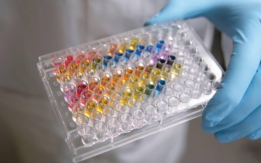
Once the cells are prepared and loaded with the indicator, they are placed in a microplate reader or under a high-resolution imaging system. When a test compound or a known agonist is added, the resulting change in fluorescence intensity is measured. This change is directly proportional to the amount of calcium being released or entering the cell. It is a highly sensitive method that allows for the detection of even subtle shifts in cellular behaviour.
Key components of a successful assay
- Fluorescent Indicators: Choosing the right dye, such as Fluo-4 or Fura-2, is critical for the sensitivity and timing of the experiment.
- Cell Lines: Stable cell lines expressing the target of interest, such as G protein-coupled receptors (GPCRs), are essential for reproducible results.
- Buffer Systems: Maintaining the correct pH and physiological conditions ensures that the cells remain healthy and responsive throughout the procedure.
- Detection Equipment: High-throughput plate readers equipped with liquid handling capabilities allow for the simultaneous addition of compounds and measurement of response.
Why this assay remains a staple in drug discovery
In the pharmaceutical industry, the search for new medicines often involves screening thousands, if not millions, of compounds to find those that interact with specific biological targets. The calcium flux assay is particularly well-suited for this task because it is fast, scalable, and provides functional data. Unlike simple binding assays that only tell you if a drug sticks to a receptor, a calcium assay tells you if the drug actually does something to the cell.
The majority of drugs on the market today target GPCRs, a massive family of receptors involved in everything from sight and smell to heart rate and mood regulation. When a GPCR is activated, it often triggers a signalling cascade that leads to a calcium flux. By using a calcium flux assay, researchers can quickly identify whether a new drug candidate acts as an agonist (activating the receptor) or an antagonist (blocking the receptor).
Optimising the assay for better data quality
While the concept is straightforward, getting high-quality, reliable data requires careful optimisation. One of the biggest challenges in these experiments is background noise. If the fluorescent dye leaks out of the cells or if the test compounds themselves are naturally fluorescent, the results can be skewed. To combat this, researchers often use ‘quenchers’ or specific wash steps to remove extracellular dye, ensuring that the signal being measured is truly coming from inside the cell.
Another factor to consider is the ‘sampling rate’. Calcium signals can be incredibly fast, peaking within seconds of stimulation. If the plate reader is too slow, the peak might be missed entirely. Modern high-throughput screening (HTS) systems are designed to handle this by using integrated pipetting systems that add the stimulant and begin reading the fluorescence almost instantaneously.
Common applications in modern research
- High-Throughput Screening: Rapidly testing large libraries of chemical compounds to find potential leads for new drugs.
- Safety Pharmacology: Assessing whether a new drug might have off-target effects on the heart or nervous system by looking at unintended calcium movements.
- Ion Channel Research: Studying the mechanics of how calcium-permeable channels open and close in response to electrical or chemical signals.
- Secondary Messenger Analysis: Mapping out complex intracellular pathways to understand how different signals are integrated within a single cell.
The shift towards more complex cell models
Historically, many of these assays were performed using immortalised cell lines like HEK293 or CHO cells because they are easy to grow and manipulate. However, there is a growing trend towards using more physiologically relevant models, such as human induced pluripotent stem cell (iPSC)-derived cardiomyocytes or neurons. These cells behave much more like the cells in a living human body, providing data that is more likely to translate into clinical success.
In cardiac research, for example, the calcium flux is what drives the rhythmic contraction of the heart. By performing a calcium flux assay on iPSC-derived heart cells, scientists can observe the ‘calcium transient’—the rise and fall of calcium that occurs with every single heartbeat. This allows them to see if a drug causes arrhythmias or other dangerous side effects long before it is ever tested in a human patient. This level of detail is revolutionising how we approach drug safety and efficacy.
Technological advancements and the future of the assay
Technology continues to push the boundaries of what is possible with calcium imaging. We are seeing the development of genetically encoded calcium indicators (GECIs), which are proteins that the cell produces itself. This eliminates the need to load chemical dyes and allows for long-term monitoring of calcium levels over days or even weeks. This is particularly useful for studying chronic drug effects or developmental processes.
Furthermore, improvements in microfluidics and ‘organ-on-a-chip’ technology are allowing researchers to perform these assays in environments that mimic the complex structures of human organs. By combining these advanced models with sensitive calcium detection, the industry is moving closer to a future where drug discovery is faster, more accurate, and less reliant on animal testing. The ability to organise and interpret the massive amounts of data generated by these high-speed assays is also being transformed by machine learning, which can identify patterns in the calcium signal that might be invisible to the human eye.
The versatility of the technique means it can be adapted for almost any area of biology. From understanding how the brain processes information to finding new ways to treat metabolic disorders, the study of calcium movement remains at the heart of scientific discovery. As we refine our methods and develop more sensitive tools, our understanding of these intricate cellular dances will only continue to grow, paving the way for the next generation of medical breakthroughs.

Marie focuses on mixed media and digital art, highlighting evolving practices and artistic innovation.